Anatomy and Physiology: Can You Hear Me Now?
Can You Hear Me Now?
There is an elegant, mechanical simplicity to sound. Sound is basically based on vibrations. Vibrating objects produce vibrations, basically waves of compression, in the air that is in contact with those objects. The vibrating skin of a drum will thus produce more sound than a rigid piece of cement struck with the same stick.
The vibration of objects helps explain why you can hear a vibrating tuning fork that is touching the top of your head, because the sound travels through vibrations in your cranial bones. As a kid, did you ever make a “phone” out of two paper cups and a string? For the phone to work, the string needs to be taut, because the vibrations must travel along the string; the two paper cups function to focus the vibrations onto the string on one end, and to amplify the vibrations from the string on the other end.
This, in a nutshell, is what happens (remember to keep the string taut):
- When you contract the muscles of your larynx (see The Respitory System), the exhaled air causes vibrations in the vocal folds.
- Vibrations in the vocal folds cause vibrations in the air, which are amplified through the resonating chambers of our paranasal sinuses.
- The vibrations in the air molecules cause the cup on the speaking end to vibrate.
- Vibrations in the cup cause the string to vibrate.
- Vibrations in the string cause the cup on the listening end to vibrate.
- Vibrations in the cup cause the waves of compression known as sound waves at the same frequency as the original vibration!
As you will see, what happens in our ear is really just more of the same!
Not Just Corn
Medical Records
I talked about the ceruminous glands in the ear canal that produce wax to keep objects and critters away from our eardrum. What you might not know is that an overproduction of wax, and subsequent buildup, can result in a blockage that makes it extremely difficult to hear. Due to the danger of puncturing one's eardrum, it is wise to leave the cleaning to a medical professional experienced in such matters! That's why doctors say the only thing you should clean your ear with is your elbow!
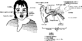
In many ways, the ear is little more than a slightly more sophisticated string-and-cup “phone.” The shape of the outer ear, called the pinna (the outer edge is the hilus, and the bottom is the lobule), captures the sound waves and amplifies them. If you have never tried it, try cupping your hand behind your ear when you listen to music, or to someone talking, and you will notice how much that amplifies the sound.
This is the principle behind the original, needless to say old-fashioned, horn-shaped hearing aids. It also explains why rabbits evolved such big ears: the better to hear predators. (Big ears, however, can also be used to radiate excess body heat.) The ear canal, or external auditory meatus, is also involved in the amplification and transmission of sound, serving to focus it on the next stop, the eardrum, which is analogous to the first cup in our cup-and-string “phone.”
Good Vibrations
The eardrum is also called the tympanic membrane. As you can imagine, its role is to receive the vibrations focused by the ear canal. Amplifying the vibrations even further requires a little help. Remember the string in our “phone” from earlier? Well, there are three little bones that do the job of the string here, three bones that students often forget when building a list of all 206 bones: the auditory ossicles. These “little bones” are, in order from the eardrum inward, the malleus (“hammer”), the incus (“anvil”), and the stapes (“stirrup”).
Medical Records
You've probably noticed the connection between the ear and the throat if you have ever had your ears plugged. By swallowing you lower the air pressure in your throat, thus equalizing the pressure at both ends of the Eustachian tube (pop!).
Just like any other bones with movable joints, there are ligaments involved. Apart from the articular cartilage between the bones, there are two ligaments—a superior one and a lateral one, each of which attaches the malleus to the inside wall of inner ear (in the temporal bone)—that help to stabilize the malleus as a result of vibrations in the eardrum. The auditory ossicles further amplify the sound. The end of the stapes attaches to the oval window of the cochlea. This connection transfers the vibration from the ossicles to the cochlear fluid. Figure 21.3 shows the parts of the ear that are involved in the transmission and amplification of sound.
At this point it is helpful to know about the other lesser-known round window. When the vibrations make their way through the coil of the cochlea, the round window's thin membrane allows the vibrations to escape. Without it, the vibrations would bounce around the cochlea until they eventually lost energy, which would sound a bit like a fast-repeating echo. The round window is right near the opening of the auditory tube, or Eustachian tube, which is where the vibrations, now transferred to the air in the inner ear, finally escape to the throat.
Hair Cells
Now that the vibrations are in the cochlear fluid, how does that translate to nerve impulses? The cochlea is a snail-like coil (membranous labyrinth) filled with a fluid called perilymph, which is more like cerebrospinal fluid than lymph. Connected to the cochlear branch of the vestibulocochlear nerve (N VIII), this is the part of the inner ear that receives the vibrations from the auditory ossicles.

Figure 21.3The structures of the ear are primarily involved in the transmission and amplification of sound. (LifeART©1989-2001, Lippincott Williams & Wilkins)
There are two regions called the scala tympani, that contain the perilymph, but sandwiched between the two, along the outer edge of the cochlea, is the spiral cochlear duct (scala media), which is filled with endolymph (similar to the fluid inside cells). Within this duct is a gelatinous glycoprotein layer called the tectorial membrane, which stimulates hair cells that lie beneath it (see Figure 21.4). As we shall see, these hair cells are found in the part of the ear that is responsible for our sensation of balance.

Figure 21.4The friction of the hair cells against the tectorial membrane stimulates the neurons of the cochlear nerve. (LifeART©1989-2001, Lippincott Williams & Wilkins)
Inside the cochlear duct is a spiral organ of Corti with multiple rows of hair cells (with supporting cells) that lie just above the basilar membrane that separates the cochlear duct from the scala tympani. These hair cells have anywhere from 50 to 100 (depending on their location) microvilli that are surrounded by endolymph. The tips of these microvilli touch the bottom of the tectorial membrane.
When the vibrations of the stapes are transferred into pressure waves in the perilymph of the scala tympani, the vestibular membrane (on the upper portion of cochlear duct, opposite the basilar membrane) starts to vibrate. This vibration creates pressure waves in the endolymph, causing movement of the tectorial membrane. The movement of the tectorial membrane creates contact with the microvilli of the hair cells, which opens up ions on the basal ends, which are attached to the cochlear branch of the vestibulocochlear nerve (N VIII). Sound!

Excerpted from The Complete Idiot's Guide to Anatomy and Physiology © 2004 by Michael J. Vieira Lazaroff. All rights reserved including the right of reproduction in whole or in part in any form. Used by arrangement with Alpha Books, a member of Penguin Group (USA) Inc.
To order this book direct from the publisher, visit the Penguin USA website or call 1-800-253-6476. You can also purchase this book at Amazon.com and Barnes & Noble.
