Anatomy and Physiology: The Four Chambers
The Four Chambers
So why do you need four chambers if three worked just fine for frogs and lizards? Humans, and indeed all mammals (not to mention birds!), are endothermic (warm blooded). Warm bloodedness requires a great deal of oxygen, for the oxygen is used to generate both ATP and heat. A four-chambered heart is an enormous evolutionary advantage over a three-chambered heart. To understand this, you need to look at the chambers and the circuits together.
Medical Records
Some babies are born with a ventricular septal defect, which means an opening between the left and right ventricles, which means that their hearts are acting like three-chambered hearts. Surgery to correct the defect is necessary in order for the child to live a normal life.
Remember the fish, with an atrium to receive blood from the body, and a ventricle to pump it out again? Well, with a three-chambered heart there are two ventricles and one atrium. The two atria emphasize a higher degree of separation between two of the circuits: the pulmonary circuit and the systemic circuit. At this point you need to start thinking of the heart in terms of left and right. The right atrium receives deoxygenated blood (low in O2, and high in CO2) from the systemic circuit, and the left atrium receives oxygenated blood (high in O2, and low in CO2) from the pulmonary circuit.
Medical Records
Don't forget that left and right in all these discussions always means the patient's left and right, which means you need to pay attention to whether any diagrams are in anterior or posterior view!
This advance was only so good, however, because both atria pump the blood to the single ventricle. In a three-chambered heart the blood pumped out of the ventricle is a mixture of both oxygenated and deoxygenated blood. This blood is pumped out to both the pulmonary and the systemic circuit (in truth, because it is pumped right back to the tissues of the heart, it really goes to all three circuits). For ectothermic (cold blooded) animals that is plenty of oxygen, but it's just not enough for you.
Birds and mammals evolved a ventricular septum, turning one ventricle into two. The result is the evolution of entirely separate pulmonary and systemic circuits (see Figure 11.2). The blood sent to the lungs is completely deoxygenated, and the blood pumped out to the rest of the body is fully oxygenated. The evolution of two ventricles, making a four-chambered heart, doubled the amount of O2 being sent to the tissues. The amount of food and waste in the blood going to the systemic circuit is not so cut and dried (see Cardiovascular and Lymphatic Circulation).
In the human heart the right atrium sends deoxygenated blood from the body to the right ventricle, which then pumps it to the lungs (pulmonary circuit). The left atrium sends oxygenated blood from the lungs to the left ventricle, which then pumps it to the body (systemic circuit).
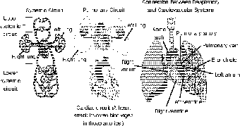
Figure 11.2The human heart has four chambers, which equally separate the right and left sides of the heart, maximizing the oxygen content of the blood being sent to the systemic circuit. (LifeART©1989-2001, Lippincott Williams & Wilkins)
Blood Vessels and Chambers
When you look at the orientation of the heart at the bottom of the thoracic cavity (see The Respitory System to learn about the pericardium) you will see that, rather than being straight up and down, the heart is at an angle, and a bit twisted (kind of like me!). This is due in part to making room for the liver, and in part to the location of the many blood vessels that attach to the heart.
Figure 11.2 shows the blood vessels connected to the heart, but you may find the flowchart in Figure 11.3 a bit easier to understand. Don't forget that the blood flow in the pulmonary and systemic circuits is continuous, meaning that blood from one circuit moves on immediately to the other circuit. Next, the central location of the heart means that blood going to the lungs needs to be pumped both left and right, and blood going to the body needs to be pumped both up and down. Thinking in terms of opposites will help you to remember the vessels.

Figure 11.3This flowchart illustrates the flow of blood, in terms of opposite directions, to and from both the systemic and pulmonary circuit. (©Michael J. Vieira Lazaroff)
Remember, there is no particular place where all of this starts, given that the circuits are continuous. Let's start with the oxygenated blood in the arteries of the systemic circuit, leaving the left ventricle of the heart via the aorta. Immediately after leaving the top of the heart, all blood vessels enter and leave through the top of the heart, the aorta arches downward to send blood to the lower body. At the top of the arch there are three large branches that go to the upper body; in this way, the systemic circuit is divided in two.
Crash Cart
A common mistake is to define arteries as vessels carrying oxygenated blood, and veins as vessels carrying deoxygenated blood. Although this is generally true, there are two important exceptions, because of the true definitions of arteries carrying blood away, and veins carrying blood to the heart. The two exceptions, which make perfect sense, both involve the pulmonary circuit: The pulmonary arteries carry deoxygenated blood to the lungs to be oxygenated, and the pulmonary veins carry the newly oxygenated blood away from the heart!
After picking up and delivering various materials in the capillaries of the upper and lower body, becoming deoxygenated in the process, the veins drain into the largest veins in the body, the superior vena cava and the inferior vena cava. Anyone who works with quadrupedal animals should know that those same vessels are called the anterior and posterior venae cavae (plural for vena cava). The venae cavae drain into the upper and lower portions of the right atrium; since the right atrium is in the upper third of the heart, this inferior vena cava is still considered attached to the top of the heart.
As the right atrium contracts, the blood must pass through a valve between the atrium and the ventricle. This valve is called the tricuspid valve (for its three cusps or flaps), or the right atrioventricular (AV) valve. Once the blood is pumped out of the right ventricle, the right AV valve prevents backflow into the right atrium. The contraction of the right ventricle does pump the blood through another valve, the pulmonary semilunar valve (named for its half moon shape), and into the pulmonary trunk. Just as the aorta splits, so does the pulmonary trunk, but this time the blood splits into the left and right pulmonary artery, in order to go to both lungs. (To see what happens next, take a deep breath and read up on the respiratory system in The Respitory System.)
Flex Your Muscles
A good way to remember the difference between the two atrioventricular valves—the tricuspid (right AV valve) and the bicuspid (left AV valve)—is to think about the dissolved gases in the blood as it passes through those valves. The deoxygenated blood passing through the tricuspid valve contains CO2, which contains three atoms (tri = three), and the oxygenated blood passing through the bicuspid valve contains O2, which contains two atoms (bi = two). A pretty cool coincidence, considering the valves were named because of their structure!
Blood returning to the heart always returns from separate vessels, whereas blood leaving the heart always leaves from a single vessel and then splits to go in opposite directions. Having vessels in pairs makes sense, but single vessels leaving the heart? Why? Think about the shape of the heart. The cone shape of the apex gives a hint about the way the heart contracts. The contraction of the ventricles, which happens simultaneously, narrows the lumen of the ventricles, as well as shortening the length of the ventricles, which pumps the blood up! It is more efficient, in ensuring the equal flow to both lungs, for example, to have the blood leave one vessel, only to split later.
Oxygenated blood returns from the two lungs through the pulmonary veins, which attach to opposite sides of the left atrium. The rest of the trip is almost the same as on the right side: the left atrium pumps the blood through the left AV valve (or bicuspid valve) into the left ventricle, and the ventricle pumps the blood through the aortic semilunar valve into the aorta.
Just as the ventricular walls are thicker than the atrial walls (because of the difference in the distance the blood is pumped), the left ventricle, which has to pump to the entire body, has thicker walls than the right ventricle, which pumps blood only to the neighboring lungs. The thick left ventricular walls also provide a greater pressure on the left AV valve with each ventricular contraction. This valve, also called the mitral valve, can sometimes bulge into the left atrium, which is called mitral valve prolapse.
To help prevent such prolapses, there are fibrous, tendon-like cords called chordae tendineae. These connective tissue cords support the valve whenever the ventricles contract. Every time a ventricle contracts, there must be enough pressure in the contraction to exceed the pressure in the pulmonary trunk or the aorta, and thus push through the semilunar valves. This puts a great strain on the AV valves, so in addition to the chordae tendineae, there are small muscles attached to the bottom of the chordae tendineae, called papillary muscles, that contract whenever the ventricles contract.
So just one question: why are there no valves where the blood enters the atria? There are two reasons for this. The first is that the blood in veins returning to the heart is at extremely low pressure, so low that it could not easily push through the closed valves already in the veins. The other reason involves the weaker contraction of the atria. The atria contract when the ventricles are relaxed, which means that the lower pressure of the ventricles at that point will make it easier for the blood to flow in that direction than backward into the veins that are filled with blood.

Excerpted from The Complete Idiot's Guide to Anatomy and Physiology © 2004 by Michael J. Vieira Lazaroff. All rights reserved including the right of reproduction in whole or in part in any form. Used by arrangement with Alpha Books, a member of Penguin Group (USA) Inc.
To order this book direct from the publisher, visit the Penguin USA website or call 1-800-253-6476. You can also purchase this book at Amazon.com and Barnes & Noble.