Anatomy and Physiology: Gyri and Sulci
Gyri and Sulci
One of the things that makes a brain instantly recognizable is the shape of its surface. The long, meandering hills (gyri, singular = gyrus) and valleys (sulci, singular = sulcus) provide a greater surface area to the brain. In general, the greater the surface area of the brain, the larger the amount of the brain that is active, and the more neurons in those areas. You can see, simply by measuring the skulls of fossil hominids (our prehuman ancestors), that the cranial cavity encasing the skull has evolved to be larger. By comparing the brains of other species, you can see that the human brain has far more, and more pronounced, gyri and sulci than more primitive mammals. This means that, although the entire brain has evolved and become larger, the cerebral hemispheres, (in particular the gray matter of the cerebral cortex) have evolved faster, as mental abilities became more important to our survival than physical ones. Think of it this way: The brain's surface area is 2200 cm2 (or 2.5 ft2), which can only fit in our skull by folding in on itself. As the brain grows and develops, the gray matter grows much faster than the white matter, which forces it to form gyri and sulci as it folds in on itself.
Also, in terms of development, it is helpful to know about the different areas of the brain and brainstem. The developing brain only has three parts, or vesicles, by the end of the third week of life. The parts of this primary brain are as follows, from the spinal cord up: rhombencephalon (“hindbrain”), mesencephalon (“midbrain”), and prosencephalon (“forebrain”). The following table traces the development of each of those three sections.
| The Development of the Areas of the Brain | |||
|---|---|---|---|
| Primary Brain Vesicles (3 Weeks) | Secondary Brain Vesicles (6 Weeks) | Brain Areas at Birth (40 Weeks) | Consist of These Areas |
| Prosencephalon | Telencephalon diencephalon | Cerebrum diencephalon | Cerebrum, thalamus, hypothalamus |
| Mesencephalon | Mesencephalon | Mesencephalon | Mesencephalon |
| Rhombencephalon | Metencephalon | Cerebellum, pons, medulla oblongata | Cerebellum, pons, medulla oblongata |
| Myelencephalon | |||
CSF and Ventricles
The CSF, or cerebrospinal fluid (removed in spinal taps), has three basic functions in the CNS: to cushion the CNS, to support the CNS (literally making the brain and spinal fluid buoyant!), and to act as an additional transport system between the CNS and the body.
As you can see from Figure 20.3, the CNS is constantly flowing around the CNS. This flow includes movement through each of the four ventricles: the two lateral ventricles (above and on either side of the thalamus), the third ventricle (running up the middle of the thalamus), and the fourth ventricle (between the pons and the cerebellum). The CSF in the fourth ventricle continues down the middle of the spinal cord in the central canal. The CSF also flows around the meninges in the following dural sinuses: superior and inferior sinuses (located above and inside the dural fold between the two hemispheres known as the falx cerebri), and in the transverse sinus (tentorium cerebelli, which is analogous to the bony process in the skull of the cat, between the cerebellum and the occipital bone).
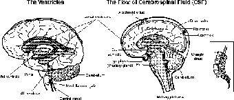
Figure 20.3The flow of cerebrospinal fluid (CSF) in and around the central nervous system (CNS). (LifeART©1989-2001, Lippincott Williams & Wilkins)
Remember those ependymal cells lining the ventricles (see The Central and Peripheral Nervous Systems)? Well, they are permeable to the CSF, thus making the CSF and the interstitial fluid in the brain and spinal cord chemically connected. This allows a spinal tap to measure the chemical health of the CNS because of chemicals that travel into the CSF from the brain and spinal tissue. CSF can travel into the veins at the arachnoid villi (refer to Figure 20.3). The total volume of the CSF is about 150 ml, but a choroid plexus in both the third and fourth ventricles constantly produces more, for a total of 500 ml a day. This means that the CSF is replaced approximately every eight hours, which makes a spinal tap a valuable tool to monitor changes.
EEG and Brainwaves
Remember the action potential and nerve impulses from The Nervous System? If each depolarization ranges from -70 mv to +30 mv, and each brain has billions of neurons, it makes sense that all that electrical activity will generate a bioelectric field. That field can be measured, just as the electric field is measured around the heart with an EKG. Because of measuring the brain's electric field, and with cephalic meaning “head,” it makes sense that the devise is called an electroencephalogram (EEG).
There are four basic types of brainwaves. As you are reading this, your mind is concentrating on a task, which means your brain is producing high-frequency beta waves (see Figure 20.4), which it also does when you are under stress. Now, if you were to lie down and rest with your eyes closed (as is often practiced in meditation or relaxation therapy), your brain would start to produce the more relaxed alpha waves. If you were to be tapped by someone while resting, or if you heard a knock at your door, those alpha waves would run for the hills as the beta waves return.

Figure 20.4The four types of brainwaves as measured by an electroen cephalogram (EEG).
Theta waves are lower-frequency, larger waves, seen in children, and briefly during sleep in adults. If you have ever been really frustrated as an adult (who hasn't?), you have produced theta waves. What kids and frustrated adults have to do with one another, other than the former causing the latter, I can't really say …. The last type of waves are the largest, and lowest frequency delta waves, which appear during deep sleep, regardless of your age. If either theta or delta waves are seen in adults when awake, under normal circumstances, it is seen as a sign of brain damage.

Excerpted from The Complete Idiot's Guide to Anatomy and Physiology © 2004 by Michael J. Vieira Lazaroff. All rights reserved including the right of reproduction in whole or in part in any form. Used by arrangement with Alpha Books, a member of Penguin Group (USA) Inc.
To order this book direct from the publisher, visit the Penguin USA website or call 1-800-253-6476. You can also purchase this book at Amazon.com and Barnes & Noble.