Anatomy and Physiology: The Muscles
The Muscles
Now that you've learned the hard part, how the muscle cells actually work, it's time to learn the muscle names. There are about 700 muscles, including both superficial and deep, too many to cover in this. Any good college-level text will provide numerous tables that provide specifics about origin, insertion, action, and motor nerves, should you need more specific information, but they can be awfully hard to interpret.
My job here is to make it all easier. All the muscle names can become a polysyllabic sea that can leave the student at sea. The names, however, are actually very simple, once you learn the principles of muscle naming, because the names often provide incredible clues as to their location and action. This section is a roadmap to help you to explore the muscular system.
Characteristics of Muscles
There are a few characteristics of muscles that I have yet to cover The Structure of the Muscles and Muscle Cells dealt mainly with the microscopic; this section deals with the larger characteristics, the macroscopic. These details are important in understanding the larger function of skeletal muscles, whereas the last section dealt, in general, with characteristics of all types of muscles.
Origin and Insertion
Skeletal muscle, true to its name, attaches to the bones of the skeleton. Such attachment is accomplished via tendons to at least two bones. But is the attachment the same for each bone? All the physical characteristics may be the same, but the function of each attachment is very different. For one thing, in order to provide controlled movement, it is important not only that the muscle moves a bone, but that the muscle is anchored while it moves the bone. Given that, there are three parts to a typical skeletal muscle: origin, belly, and insertion (see Figure 9.1).
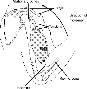
Figure 9.1Every skeletal muscle consists of tendons on either end, with Direction of an origin connected to one end, movement the insertion at the other, and the belly in the middle. (LifeART©1989-2001, Lippincott Williams & Wilkins)
The widest part of a muscle is called the belly. The origin and insertion deal with the tendon attachments. First of all, the bone needs to be anchored; the attachment to the unmoving bone is called the origin. Next, the contraction of the belly pulls on the tendons, and the other bone moves; the attachment to the moving bone is called the insertion.
The Three Classes of Levers
The origin and the insertion bring up the concept of leverage. Synovial joints not only allow for movement, but they can act as part of a lever. There are three basic parts to any lever: fulcrum (F), effort (E), and resistance (R). The fulcrum (F) is the fixed point around which the lever moves. The effort (E) is a force that causes the lever to move. Last, the resistance is the weight that is acting against the movement; this resistance includes the weight of the bone being moved, plus any object that is being carried or moved.
Levers are divided into three classes, based on the location of the three parts in relation to each other, as you can see in Figure 9.2. Their relationship to each other says quite a bit about the lever. Levers can be used to increase or decrease the amount of work done for a particular effort. The definition of work is force multiplied by distance (W = FD).

Figure 9.2The three types of levers, both in the body and in common objects. (LifeART©1989-2001, Lippincott Williams & Wilkins)
If you have ever been on a seesaw, you have been on a first-class lever (with the fulcrum in the middle). In the body, a first-class lever is used to lift your head. The effort is provided by the trapezius muscle pulling on the occipital bone, the weight of the face acts as the resistance, and the fulcrum is the joint between the occipital bone and the atlas vertebra.
Second- and third-class levers both have the fulcrum at one end; in both of these the direction of the effort is the same as the movement of the resistance. A second-class lever places the resistance in the middle; a typical example of this is a wheelbarrow. Second-class levers are the rarest in the body; one example is when you stand on tiptoe (using your gastrocnemius muscle to do plantar flexion), your toes and the ball of your foot make up the fulcrum, the effort is from the gastrocnemius, and the resistance (the weight of your body) is in the middle.
A third-class lever places the effort in the middle of the fulcrum and the resistance. These are the most common levers in the body, simply because the insertion of the muscle is proximal to the joint; were the insertion distal, there would need to be a lot of extra tissue needed to cover the muscle. (Imagine a straight muscle extending from the scapula to the distal part of the radius, and the inside of your elbow would disappear!) The disadvantage (mechanical disadvantage) of third-class levers is that quite a bit more strength is needed (effort) to pull on the bone (resistance).
Flex Your Muscles
To compare the amount of effort needed in a second- and a third-class lever, place your elbow on a table and relax your forearm completely. Now pinch your skin on the back of your hand and lift your forearm. By placing the effort far from the fulcrum (your elbow), you have made a second-class lever. Now pinch your skin close to the inside of your elbow and lift your forearm … not so easy, is it? You have just switched the location of the effort and the resistance, making a third-class lever.
Arrangement of Fascicles
Fasciculi, or fascicles (see The Structure of the Muscles and Muscle Cells), can be arranged in very different ways. In parallel muscles the fascicles are, well, parallel, and the belly is the same width as the tendons. Most people think of muscles as being fusiform, which means that the fascicles are almost parallel, but that the belly is wider, and the muscle tapes toward both the origin and the insertion. Both these types have short tendons on each end.
Pennate muscles have shorter fascicles and much longer tendons, sometimes almost as long as the whole muscle. The location of the fascicles, and the number of tendons, determines which type of pinnate muscle it is. If the fascicles are on one side, it's called unipennate, and it's bipennate if the fascicles are on both sides. Multipennate muscles have multiple tendons, with the fascicles on both sides of the tendons. Figure 9.3 shows the various possible arrangements of fascicles in muscles.
The only muscle arrangement left is circular. These muscles have the fascicles in roughly concentric circles around an opening. The orbicularis oris, around the mouth (pucker up!), and the orbicularis oculi, around the eyes (Wink! Wink!), are examples of this type.

Excerpted from The Complete Idiot's Guide to Anatomy and Physiology © 2004 by Michael J. Vieira Lazaroff. All rights reserved including the right of reproduction in whole or in part in any form. Used by arrangement with Alpha Books, a member of Penguin Group (USA) Inc.
To order this book direct from the publisher, visit the Penguin USA website or call 1-800-253-6476. You can also purchase this book at Amazon.com and Barnes & Noble.