Anatomy and Physiology: Neurons
Neurons
Okay, slow down. Axons? Dendrites? Cell bodies? Remember, neural tissue is important in its ability to communicate from one region of the body to another. Unlike the endocrine system which communicates slowly, using hormones carried by the cardiovascular system, neurons must be able to communicate quickly! The only way to ensure this is to make actual physical, cellular connections between different areas of the body. Neurons are designed to carry nerve impulses, basically waves of depolarization (see The Structure of the Muscles and Muscle Cells “You've Got Potential”), along the length of the cell, which is made up of cytoplasmic extensions called dendrites and axons.
The Parts, To and From
Flex Your Muscles
The word perikaryon should sound a bit familiar. Remember learning about prokaryotic and eukaryotic cells? The word karyon means kernel and refers to the nucleus. Prokaryotic cells are cells that evolved before (pro as in prologue) a nucleus, and eukaryotic cells have a good (eu as in euphemism) nucleus. Well, in the same sense, the perikaryon or cell body is the area around (peri as in perimeter) the nucleus.
When everyone thinks about typical human cells, they probably see a cell with a nucleus in their mind (unless they are thinking about red blood cells!). Neurons, too, have a nucleus, but it is away from where the action is! The most “normal-looking” part of the cell is the cell body, perikaryon, or soma, which contains the nucleus. Although the nucleus is usually a hotbed of activity, it is less so in neurons. Very little cellular activity actually happens here, which is one of the reasons the supporting work of the neuroglia is so necessary. Most neurons lack centrioles in their somas, which is the other reason why scientists believed for so long that adult neurons didn't divide.
On either side of the cell body are the parts of the neuron that make it famous: the dendrite and the axon. The difference between the two is basically functional, in that it is not always possible to tell which is which under the microscope. The function of the dendrite is to carry the nerve impulse to the cell body, and the axon carries the impulse away from the cell body (see Figure 19.3). The basic nature of a neuron, unlike a phone cord, or a fiber-optic cable, involves only one-way transmission. So the transmission is always as follows: dendrites, cell body, axon.
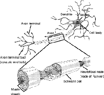
Figure 19.3The dendrites and axons of a neuron carry neural impulses to and from the cell body. (LifeART©1989-2001, Lippincott Williams & Wilkins)
Medical Records
Viruses and toxins have been known to take advantage of fast axonal transport by hitching a ride. The toxin from tetanus bacteria, whose effect led to the name lockjaw, is one such culprit. The herpes virus is another culprit; herpes zoster, which causes chicken pox, is extremely painful if and when it reoccurs later in life as shingles, because it attacks nerves, traveling through the nerve root from the dorsal root ganglion. Lastly, the rabies virus will work its way to the brain, and the speed of the transport means a bite on the arm or neck requires earlier medical intervention than a bite on the leg!
Dendrites are usually unmyelinated (thus making dendrites part of the gray matter), whereas axons can be either myelinated (white matter) or unmyelinated. The real action takes place both along the dendrites and axon (the nerve impulse) and at the connection between the neuron and other neurons, or neurons and muscle cells, for example; these connections are called synapses (see The Structure of the Muscles and Muscle Cells). Synapses always involve the end of axons, called axon terminal bulbs, or synaptic end bulbs. Although some of the neuron to neuron synapses occur at the perikaryon, the majority of them are found at the dendrites, which account for 80 to 90 percent of the surface area of your garden variety neuron.
Those long axons and dendrites don't sound very sturdy. Sure the oligodendrocytes and Schwann cells must help, as well as the astrocytes, but isn't there any inner framework? I'm glad you asked! The cytoskeleton is alive and well here, but traveling incognito. The microtubules and microfilaments go by the names neurotubules and neurofilaments here; together they are bundled as neurofibrils, which run the length of the dendrites and axons, providing internal support.
Axons have another trick up their sleeve! They have their own inner transport mechanisms, which differ from the rapid membrane-based nerve impulses. Slow axonal transport involves the movement of chemicals along the axoplasm (axonal cytoplasm); this cumbersome method only goes one way, carrying chemicals to the axon terminals at the rate of a whopping 1 to 5 mm a day (ooooh!). Fast axonal transport is 40 to 400 times faster, and has the advantage of going both ways (anterograde = from the soma; retrograde = to the soma) via the neurotubules in the axon. In addition to molecular cargo, organelles and even viruses have been known to take a ride on the fast axonal transport.
The Insulated Wire
Remember those pesky neurolemmocytes? Well, these Schwann cells, as I said earlier, insulate the axon. The insulation is very much like the insulation on an electric wire. If you have ever done projects with electricity in school, then you might have noticed that electricity running through coiled wire exerts an electromagnetic field. You can't see the field, of course, but simply watching a compass needle jump shows that it is there.
This field indicates the movement of electrons away from the wire. The insulation keeps the electrons traveling along the wire, thus eliminating “leakage.” In the same sense, the movement of Na+ and K+ ions in and out of neurilemma is easier if there isn't too much area for them to enter and leave.
The analogy gets tricky if you take it too literally, because the transmission of electricity is along the wire, whereas the transmission along the axon takes place on the outside of the “wire.” The key is the spaces between the Schwann cells. These little gaps, connected by little sections of bare axons, are called neurofibral nodes, or nodes of Ranvier. For this reason, Schwann cells, and their myelin sheath, are also called myelinated internodes.
Simply calling these cells myelinated internodes illustrates the importance of the nodes. Remember the whole process of polarization and depolarization of membranes in muscle cells (see The Structure of the Muscles and Muscle Cells)? Energy is required to power the active transport that makes a membrane polarized. Depolarization happens fast due to facilitated diffusion. The difference here is that, unlike the wave of depolarization along a myofiber (muscle cell), the depolarization jumps from node to node. This process, known as saltatory conduction (as opposed to continuous conduction in unmyelinated axons) helps to speed up the propagation of the nerve impulse.

Excerpted from The Complete Idiot's Guide to Anatomy and Physiology © 2004 by Michael J. Vieira Lazaroff. All rights reserved including the right of reproduction in whole or in part in any form. Used by arrangement with Alpha Books, a member of Penguin Group (USA) Inc.
To order this book direct from the publisher, visit the Penguin USA website or call 1-800-253-6476. You can also purchase this book at Amazon.com and Barnes & Noble.