Anatomy and Physiology: Support Staff
Support Staff
There is an old saying that you only use 10 percent of your brain. I've never understood this expression, for we use all of it, although not at the same time. The expression has something to do with the fact that we have far more (up to 50 times more!) support cells, called neuroglia, or glial cells, than we do neurons. Since those neuroglia perform essential functions for the neurons—including regulating ion concentration, insulating axons, and lining ventricles—it seems odd to consider them as unused! This section concentrates on the many ways neuroglia make themselves indispensable, allowing us to use 100 percent of our brain!
In the CNS
You might have heard the expression gray matter, and perhaps you equated it with the brain. But did you also know about white matter? The difference between the two depends on one of the forms of neuroglia. Each neuron has a cytoplasmic extension called an axon (see the neuron section of this section) that carries the messages away from the cell body; in order to speed up the message the axon is surrounded by an insulating sheath made of myelin. The myelin sheath is simply a phospholipid cell membrane wrapped around and around to protect the axon, similar to the way the plastic coating on an electric wire preserves the direction of the electricity, preventing it from traveling outward.
The myelin sheath is white in color and is provided by specific neuroglia (oligodendrocytes in the CNS, and neurolemmocytes or Schwann cells in the PNS). The white matter in the brain and spinal cord is due to the high concentration of myelinated axons in that area, due to the white color of the myelin. The gray matter, on the other hand, contains either unmyelinated axons or mostly dendrites and cell bodies.
The central nervous system has four types of neuroglia, as opposed to only two for the peripheral nervous system. Part of the reason for this is the basic nature of the two systems, for the PNS is basically concerned with the transfer of information to and from the CNS, but the CNS is the system that must actually make the decisions (conscious or otherwise) that regulate the rest of the body. The wider range of functions in the CNS requires a wider range of glial cells.
In addition to the oligodendrocytes mentioned so far, there are three others: ependymal cells, microglia, and astrocytes. Ependymal cells are basically epithelial cells (squamous to columnar), and they are found lining the ventricles. Microglia are pretty cool because they play the role of macrophages, cleaning out cellular waste, attacking invaders, and so on. Although they are usually fixed, they have been known to find their way to damaged tissues.
Astrocytes are a bit more complex. These cells have numerous cellular extensions, which help to maintain a framework holding together the various axons and dendrites in the CNS. In addition, this framework apparently guides the connections of the growing neurons during development. The framework, mentioned above, is also crucial in terms of repairing damaged tissues. One of the most interesting parts of their job is to help provide what is called the blood-brain barrier (BBB).
Blood-Brain Barrier
Bizarre as it might seem, there is in fact a difference between the function of capillaries in the brain and those in the rest of the body. One of the reasons for this is, well, capillaries aren't very leaky! You might remember some of the tricks of capillaries to release plasma (see Cardiovascular and Lymphatic Circulation), including gaps between the endothelial cells that make up the thin capillary walls. Brain capillaries, on the other hand, lack those gaps. They even have tight junctions to prevent leakage. Unlike most capillaries, those in the brain also have a continuous basement membrane on the outside of the endothelium. As if that weren't enough, those pesky astrocytes put their cytoplasmic projections on the outside of the capillaries (see Figure 19.2). Multiple astrocytes thus form a continuous outer layer to almost every capillary in the CNS.
The blood-brain barrier refers in part to the differential movement of materials. Certain substances pass through easily, such as glucose, O2, CO2, and H2O. As a matter of fact, insulin is not needed for glucose to leave the capillaries around nervous tissue; the high glucose levels in the nervous tissue of diabetics leads to a form of nerve damage known as neuropathy. Lipid-soluble molecules, as you would expect, travel through quite easily; this is a good thing in the case of anesthetics, but it doesn't help in terms of some rather addictive substances that also make their way through easily (alcohol, caffeine, heroin, and nicotine). Ions such as Na+, Cl-, and K+ (all a part of neural propagation), can pass through, but they rely on carrier-mediated transport. Proteins and most antibiotics cannot pass through the blood-brain barrier; this antibiotic barrier can be troublesome in terms of cranial infections.
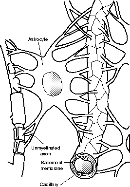
Figure 19.2The projections of astrocytes form part of the physical barrier known as the blood-brain barrier.
Not all of the brain contains such a barrier with the blood. The regions around the third ventricle, including the hypophysis, hypothalamus, and pineal gland, have normal capillary flow. The name for these regions, circumventricular organs or CVOs, should be self-explanatory (that is, “around the ventricle”). Given that these areas are involved in the monitoring of homeostasis (fluid levels, blood pressure, thirst, and hunger), it makes sense to not have the blood-brain barrier there!
In the PNS
It might come as a bit of a surprise that there is no cell equivalent to the microglia in the PNS. A closer look, however, shows that the CNS is fairly insulated from other tissues, whereas the nerves of the PNS are embedded in other tissues, particularly the nerve endings. Those tissues are already serviced either by wandering or by fixed macrophages.
There are only two types of neuroglia in the peripheral nervous system: satellite cells and neurilemmocytes, also known as Schwann cells. The satellite cells are important in terms of clumps of nerve cells known as ganglia. Ganglia contain more than just axons and dendrites, the makeup of nerves; ganglia (singular = ganglion) also contain the more cumbersome cell bodies. These cell bodies are surrounded by the flattened satellite cells, which basically provide protection.
Flex Your Muscles
The name neurolemmocyte makes sense when you remember that the cell membrane of neurons is called the neurilemma (like the sarcolemma on muscle cells). These cells wrap around the neurilemma, thus insulating it!
Remember how the axon carries messages away from the cell body? Well, the long axons of the PNS require insulation just as much, if not more so, than in the CNS. This insulation is provided by neurilemmocytes. These weird little pill-shaped cells grow around a small section of an axon, gradually producing more and more membrane, which ends up wrapping layer after layer of tightly coiled membrane.
These multiple layers of phospholipid bilayers form the myelin sheath. The white color of the myelin sheath is what gives myelinated nerve tissue the name white matter; gray matter, on the other hand, is made up of either unmyelinated axons, or the dendrites and cell bodies of neurons. It is important to note here that all axons in the PNS are protected from the interstitial fluid by neurilemmocytes, but that these Schwann cells don't always wrap around the axon a freakishly large number of times. When each Schwann cell wraps around a single axon multiple times, the axon is myelinated. When each Schwann cell wraps around multiple axons, but only once around each one, then the axons are not myelinated.

Excerpted from The Complete Idiot's Guide to Anatomy and Physiology © 2004 by Michael J. Vieira Lazaroff. All rights reserved including the right of reproduction in whole or in part in any form. Used by arrangement with Alpha Books, a member of Penguin Group (USA) Inc.
To order this book direct from the publisher, visit the Penguin USA website or call 1-800-253-6476. You can also purchase this book at Amazon.com and Barnes & Noble.