Anatomy and Physiology: Cells, Bells!
Cells, Bells!
My first introduction to white blood cells was that old sci-fi classic, Fantastic Voyage. (I recommend it, but to have the most fun, you should keep an eye out for the mistakes!) One of the final scenes involves the bad guy being eaten by a white blood cell! You may not have learned, however, that while there is only one type of red blood cell (RBC), that there are many white blood cells (WBCs), not to mention platelets! All of these cells are called formed elements, and they make up about 45 percent of blood volume.
Medical Records
Note that any percentages given for substances in the blood, either within the plasma, or as part of the formed elements, are averages. Your health may affect the amount of each substance. A drop in the number of erythrocytes (RBCs), for example, is one form of anemia. For this reason, blood tests can be very revealing. As a diabetic, I test my blood glucose level with a handheld meter up to eight times a day.
In The Bones, I mentioned that the red bone marrow was the site of blood cell production in a process called hemopoiesis, which is summarized in Figure 10.1. In truth, not all blood cells are made in the red bone marrow, because while the B-lymphocytes are made in the bone (B for bone), the T-lymphocytes are made in the thymus (T for thymus). All of the blood cells come from the same stem cell—the pluripotent hematopoietic stem cell, its friends call it a hemocytoblast—in a model of differentiation.
This figure may be a bit frightening, but try to look for the commonalities. From the one stem cell (the hemocytoblast) there are five cells into which it can develop. Note that the first cells all have the suffix “-blast,” and that the three of the five cells have, in their name, a clue as to the final cell (for example, a lymphoblast will ultimately become a lymphocyte). The second step, with the exception of the early erythrocyte, all have similar names (for example, promonocytes ultimately become monocytes). Look for such strands as you follow the diagram, and you will see that it is not so frightening after all!
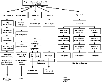
Figure 10.1This diagram shows the development of all types of blood cells from the original hemocytoblast. (©Michael J. Vieira Lazaroff)
Now I Know My RBCs
RBCs make up 99.9 percent of all formed elements, that's a nearly a 1000 to 1 ratio! RBCs give blood its red color. I always tell my students that erythrocytes, or red blood cells (RBCs), are on a suicide mission. From the earlier diagram you may remember that the last step of RBC formation is the loss of the nucleus (as well as ribosomes, mitochondria, and most of the other cell organelles). That it is a very important event, for it says a tremendous amount about that cell.
Crash Cart
Guess what makes deoxygenated blood blue? Give up? Nothing! Once and for all, blood is never blue! Not even among aristocrats! Deoxygenated blood is dark red, and oxygenated blood is bright red, but it's always red! In terms of bleeding, blood cannot immediately turn red when exposed to the air, simply because of the low SA/V. Arteries are too deep to see, but veins look blue simply because the red is refracted by the tissue layers the light must pass through before reaching your eyes. As my wife says, “Blood never gets the blues!”
Without a nucleus (and its DNA cargo) or ribosomes, an RBC cannot make protein, and so it is trapped with its current complement of proteins. That means it can do very little to react to its environment, and it is completely incapable of repairing itself. The loss of mitochondria means it can process glucose only without oxygen (an irony, considering how much oxygen is in the RBC): Producing only 2 ATP versus 36, the RBC must have very low energy needs.
An RBC's role is basically as a floating sack of hemoglobin, which is the protein that carries oxygen. The iron in the hemoglobin causes the cells to turn more red in the presence of oxygen, just the way rusting iron turns red! Without a nucleus, RBCs are incapable of dividing, so they are on a suicide mission, working until they are destroyed by phagocytic cells, or in the spleen.

Figure 10.2The quaternary structure of the oxygen-carrying hemoglobin molecule. (LifeART©1989-2001, Chain 1 Lippincott Williams & Wilkins)
RBCs are a beautiful example of differentiation. Here we have a cell that does only one thing, lives for 120 days before it is broken down, and must constantly be replaced. The single-minded purpose of the cell is linked to the hemoglobin, or Hb, which makes up 95 percent of the proteins in the cell. The lack of the nucleus allows room for more hemoglobin molecules, which means that the cell can carry more oxygen.

Figure 10.3The shape of a red blood cell (RBC). (LifeART©1989-2001, Lippincott Williams & Wilkins)
So what's the big deal? After all, why can't we just carry the O2 in the plasma, as we do the other molecules? The answer is due in part to the fact that O2 is very poorly soluble in water (1.5 percent), and in part to the structure of the hemoglobin molecule itself. I discussed the quaternary structure of proteins. A hemoglobin molecule is roughly x-shaped, with two alpha chains (each at a diagonal), and two beta chains (each also at a diagonal); each of these four chains is a tertiary structure (see Figure 10.2).
The Big Picture
RBCs are mostly concerned with carrying oxygen. The need for O2 is connected to energy capture from glucose. With O2 the energy available is 19 times higher (38 ATP instead of 2). It is clear that O2 is essential for our survival, which explains the special connection between the cardiovascular and respiratory systems, as you shall see in The Heart.
In the midst of each chain is a heme group, which contains an iron ion (Fe2+). Each heme group can hold one O2 molecule, so each Hb molecule therefore carries four oxygen molecules; all this adds up to a tremendous amount considering we have about 280 million Hb molecules in every RBC! This arrangement allows the hemoglobin in the RBC to be far more efficient at carrying oxygen than plasma. In addition to carrying about 98.5 percent of the O2, hemoglobin carries about 23 percent of the CO2; the combination of CO2 and Hb is called carbaminohemoglobin. The remaining CO2 is carried as ions (see The Digestive System) in the plasma, or the cytoplasm of the RBC.
Do you remember surface area to volume ratio (SA/V)? The important idea to remember is that a smaller cell has a larger SA/V. RBC is the star of SA/V, with a SA/V of about 1.5 million to one! The shape of an RBC is a bit weird; it's shaped like a biconcave disk (refer to Figure 10.3). Think of the shape of a bagel or doughnut, but instead of a hole, the center is merely pinched in (like a bialy). This shape has a far greater SA/V ratio than a sphere of the same volume. In fact, the total surface area of all 25 trillion RBCs in the average adult's body—260 million in a single drop!—is equivalent to 3,800 m2, or 2,000 times larger than that of our skin (about 1.9 m2)!

Excerpted from The Complete Idiot's Guide to Anatomy and Physiology © 2004 by Michael J. Vieira Lazaroff. All rights reserved including the right of reproduction in whole or in part in any form. Used by arrangement with Alpha Books, a member of Penguin Group (USA) Inc.
To order this book direct from the publisher, visit the Penguin USA website or call 1-800-253-6476. You can also purchase this book at Amazon.com and Barnes & Noble.