Anatomy and Physiology: The Great Divide
The Great Divide
The nervous system can be divided two ways, anatomically and functionally. The basic anatomical division is between the central nervous system (CNS), which includes the brain and the spinal cord, and the peripheral nervous system (PNS), which includes the cranial and spinal nerves (see Figure 19.1). You might think of the spinal cord as one large nerve with many nerve branches, but as there are nerves that extend from the brainstem, and nerves extending from the spinal cord, the spinal cord is considered part of the CNS.
The job of the nervous system is basically to sense the environment and the body's place in it, coordinate either a voluntary or an involuntary response, and then carry out that response. As the true control center, the CNS needs to have access to information about the rest of the body, just like a kitchen manager in a restaurant needs to know about the attendance of the staff, the alcohol and food orders, the condition of the equipment, the reservations, and so on, in order to plan accordingly. The only way to get access to the information about the body is through the PNS.
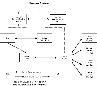
Figure 19.1The organization of the nervous system is based on the basic division between the central nervous system (CNS) and the peripheral nervous system (PNS). (©Michael J. Vieira Lazaroff)
Another way to divide the nervous system is in terms of function. In this organization of the nervous system each of the two branches includes parts of both the CNS and the PNS. The division is based on two factors: whether the response is voluntary, and the area of the body that carries out the instructions of the CNS. The voluntary portion is called the somatic nervous system (SNS), in which the responding organs are skeletal muscles. The other division is called the autonomic nervous system (ANS), which is involuntary. If you think about the organs that need to be working in the background, without having to give them thought, you know where the bulk of these organs are: in the thoracic and abdominopelvic cavities.
The Central Nervous System
The CNS is, of course, a little more complex than just “brain and spinal cord.” In looking at the brain, numerous divisions are important. In this section, I plan on only introducing the cast of characters; I will cover them in more detail in The Central and Peripheral Nervous Systems. When people usually think about the brain, their mind quickly goes to, well, the mind, or cerebral cortex, the part of the brain where thought and perception occur, but there's more.
Medical Records
It has been determined through imaging technology that women actively use both hemispheres of the brain when listening to conversations; that ol' corpus callosum is just kept jumpin'. Men, on the other hand, use mainly the left hemisphere. Is this why men aren't very good listeners?
First of all, we divide the brain into two hemispheres, with the division right along the midline. True, the either/or mentality is more a sign of the hard wiring of the mammalian brain than with our hemispheres, but the left and right hemispheres, nonetheless, do divide some of the brain's activities. Neither hemisphere works completely alone, however, for messages are passed back and forth between the hemispheres in a broad band of neurons called the corpus callosum.
The brain is divided into left and right hemispheres, with each hemisphere divided into the same lobes: frontal (anterior, of course!), parietal, temporal, and occipital. Beneath the brain is the diencephalons (made of the thalamus and the hypothalamus), which is just above the brainstem. Arising out of the top of the spinal cord, the brainstem is made of, from top to bottom, the midbrain, the pons, and the medulla oblongata. Coming out of the pons is another lobe, the cerebellum. The spinal cord, extending through all the vertebral foramina in the spinal column, continues the CNS all the way to the end of the sacrum. Flowing around the spinal cord and brain, and through the four ventricles, is the nutrient-rich protective fluid known as cerebrospinal fluid or CSF. All these aspects of the CNS will be covered in The Central and Peripheral Nervous Systems.
The Peripheral Nervous System
The brain may be the neural control center, and the spinal cord the main thoroughfare of the nervous system, but neither of these, alone, can carry messages to and from the foot soldiers, our tissues and organs. For that we need the PNS. The neurons in the PNS are constrained by the fact that every neuron can only send messages in one direction. Within the PNS, in order to do the job properly, there need to be two sets of neurons: afferent and efferent. Afferent neurons bring messages from the body to the brain; these neurons are sensory in nature, hence also being called sensory neurons. Efferent neurons, on the other hand, carry messages from the brain to the body, instructing the body as to what action to take; as the majority of these actions are made by muscle tissue (skeletal, cardiac, and smooth), these are also called motor neurons. Most peripheral nerves carry messages both ways, hence having both afferent and efferent neurons, but certain cranial nerves are limited to either sensory function, such as the optic nerve (N II), or motor function, such as the oculomotor nerve (N III).
In addition to afferent and efferent neurons, the PNS can be divided according to the type of action that the efferent nerves trigger. Those voluntary actions, which involve skeletal muscles, are part of the somatic nervous system (SNS). Muscles, however, are just the tip of the iceberg, when it comes to what actions the body can take. Look in either the thoracic or the abdominopelvic cavity, and, with the exception of the diaphragm, you'll see no skeletal muscles. Even the diaphragm is an interesting exception in that it is not entirely voluntary. It is the involuntary actions of the organs of these two cavities that led to the name of the area of the PNS that triggers them: the autonomic nervous system (ANS), although we now know that the brain regulates these functions, and that they are not truly autonomous.
The last method of dividing the PNS is purely in terms of anatomy, according to the location of the nerves. There are 12 pairs of cranial nerves, indicated as N I through N XII. A number of these are completely sensory, as so many senses are located in the head (see the following table). The sections of the spine also have their own nerves, called spinal nerves: cervical nerves (C1-C8), thoracic nerves (T1-T12), lumbar nerves (L1-L5), and sacral nerves (S1-S5).
| The Functions of the 12 Cranial Nerves | ||
|---|---|---|
| Cranial Nerve | Sensory | Motor |
| I Olfactory | Smell | (None) |
| II Optic | Vision | (None) |
| III Oculomotor | (None) | Eyeball movement |
| IV Trochlear | (None) | Eyeball movement |
| V Trigeminal | Gums, teeth, lips, facial skin, and so on | Jaw movement |
| VI Abducens | (None) | Eyeball movement |
| VII Facial | Taste (anterior 2/3 of tongue) | Facial muscles lacrimal (tear) and salivary glands |
| VIII Vestibulocochlear | Hearing/balance | (None) |
| IX Glossopharyngeal | Tongue (posterior 1/3), pharynx, palate, 02, CO2, pH, blood pressure | Pharynx, parotid salivary gland |
| X Vagus | Visceral sensation | Visceral response |
| XI Accessory | (None) | Neck, palate pharynx, larynx |
| XII Hypoglossal | (None) | Tongue |

Excerpted from The Complete Idiot's Guide to Anatomy and Physiology © 2004 by Michael J. Vieira Lazaroff. All rights reserved including the right of reproduction in whole or in part in any form. Used by arrangement with Alpha Books, a member of Penguin Group (USA) Inc.
To order this book direct from the publisher, visit the Penguin USA website or call 1-800-253-6476. You can also purchase this book at Amazon.com and Barnes & Noble.