Anatomy and Physiology: Vessels To and Fro
Vessels To and Fro
As I discussed in the previous two sections, arteries carry blood away from the heart, and veins carry blood to the heart, with capillaries doing the real work of exchange at the tissue level. The arterial and venous pathways are parallel most of the way, but that doesn't mean that arteries and veins are the same, nor are capillaries merely small arteries and veins. Their differences reflect both the pressure of the blood within the vessels, and the function of the vessels themselves.
Leaving and Returning Home
Most books are content to divide their illustrations into arteries and veins. Although they are opposite ends of the same circuit, you can best understand the nature of the blood vessels if you see them side by side, as shown in Figure 12.1.
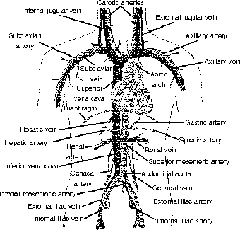
Figure 12.1This diagram shows the parallel pathways of the larger vessels in the systemic circulation. (©2003 www.clipart.com)
Several things about arteries and veins are identical. Both vessels have a thick outer layer called a tunica externa, with a layer of smooth muscle underneath called the tunica media. The hollow opening of each vessel is called its lumen, and the innermost layer, on the outer edge of the lumen is called the tunica interna, which is made of an epithelial layer (endothelium) and a basement membrane.
If you compare the structure of arteries and veins, two things immediately stand out. For one, the vessel walls are not the same; the arterial walls are much thicker than the venous walls, due in part to a larger smooth muscle layer. In addition, on either side of the muscle layer there are layers of elastic connective tissue called the internal and external elastic lamina. This layer is important due to the higher pressure the blood exerts against the arterial walls; this is, after all, what blood pressure is!
Crash Cart
If you are ever in a wedding party, don't make one of the most common mistakes of posture, for you will easily embarrass yourself! Remember the importance of the skeletal muscles in returning the blood to the heart? Well, people who lock their knees, rather than using their muscles to support their lower legs, will have blood pool in their legs, eventually leading to a critical drop in blood pressure in the arteries to the brain. With enough time, and a big enough drop in oxygen to the brain, you will faint dead away, and, unless you hit your head on the way down, the only injury will be to your pride, as long as you are forgiven for screwing up the wedding!
The other striking difference between arteries and veins is the presence of valves in the veins. Remember that the arteries are taking blood from the heart, whereas the veins are taking blood from the capillaries, so there is little pressure in the veins. Without these valves, the blood would easily pool in our legs. In addition, the contraction of our skeletal muscles in our limbs (skeletal muscle pump), as well as bellows-like pressure changes from the contraction and relaxation of our diaphragm (respiratory pump), help to pump the blood back to the heart.
Arteries, in turn, are divided into two basic types: elastic (conducting) and muscular (distributing). The elastic fibers in the conducting arteries temporarily expand and store some of the kinetic energy of the blood pumped with each heartbeat, and then transfers it back to the forward motion of the blood with each ventricular diastole (see The Heart) when the vessels recoil.
The muscular arteries are smaller, and more distant from the heart, and they are able to vasoconstrict, or contract the muscles in the vessel walls to narrow the lumen of the artery thus regulating the amount of blood to the tissues. This vasoconstriction is also carried out at the arteriole level. The relaxation of the smooth muscle around the arterioles leads to a dilation of the vessels, called vasodilation.
Arteries are probably what most people think about when they think about blood vessels. Arteries, after all, are the place where we feel our pulse, and where we measure our blood pressure. When people think of blood giving life—delivering oxygen, food, and water—they are usually thinking of arteries; it turns out, however, that things are more complicated than that. Arteries may deliver the goods, but they don't actually get off the truck until they get to the capillaries. What is even more surprising is that the highest amount of food, and the lowest amount of waste are not found in the arteries at all, but in certain specific veins (as I explain later in this section).
Pickup and Delivery
Capillaries may be small, but they do the important work! Without these unassuming little, and I mean little, vessels, none of our tissues would get oxygen, get rid of carbon dioxide and other wastes, receive nutrients, water, and so on. These capillaries connect the arterioles to the venules, but they branch extensively in the process, greatly increasing surface area to volume ratio, which is essential for all the absorption and filtration that capillaries do. (I discuss filtration and reabsorption in more detail in The Excretory System.) Diffusion is a wonderful thing, but it needs space if we are going to be able to do enough diffusion for us to survive. Remember, we are all glorified amoeba!
There are a few things that make capillaries unique. For one thing, they are incredibly small. Their lumen is so small that the tiny erythrocytes (RBCs; see The Blood) must pass through single file! This also has the benefit of helping the pickup and release of oxygen from the RBCs, since they are so close to the endothelium. The endothelium is one cell layer thick, and since they are squamous cells, they are very thin. This thin layer helps the materials to travel through the cells. On the outside of the epithelium, as always, there is a basement membrane (see The Nervous System) for attachment to the outer tissues; that basement membrane is important when vessels get damaged, for it is involved in initiating the clotting process (see The Blood).
Capillaries also are involved in vasomotion, which is the movement of blood through the capillaries via a separate pumping action. It turns out that there is a special type of vessel that travels straight through a capillary bed from an arteriole to a venule, and this vessel is called a metarteriole (see Figure 12.2). This metarteriole has smooth muscles that pump the blood, and a smooth muscle precapillary sphincter in turn contracts and relaxes, thus regulating the blood into the true capillaries. The other end of the metarteriole, which lacks smooth muscle, is a low-resistance pathway called a thoroughfare channel, and it empties into the venule. It seems that there's a whole lot of pumpin' goin' on!
What I find cool is how the capillaries, as the end stage of the closed system, manage to transfer materials. The capillaries in our muscles, connective tissue, and lungs are called continuous capillaries, and they help out the flow of materials by having occasional gaps intercellular clefts—sounds like cheating to me! Other capillaries—in the intestinal villi, kidneys, ventricles of the brain, and in endocrine glands—are called fenestrated capillaries, which means they have large pores in the endothelial cell membrane—hey, maybe this system isn't so closed after all!
As if those weren't bad enough, there are other capillaries called sinusoids, which are found in such places as the liver, spleen, and adenohypophysis (anterior pituitary), that have larger spaces between endothelial cells, plus an incomplete or missing basement membrane—what a leaky pipe that is! All of these openings, however, are only big enough for plasma and phagocytic cells; RBCs are too big and inflexible to make it through. But perhaps the coolest way of sending materials out through the endothelium is a combination of endocytosis and exocytosis. Do you remember pinocytosis? This miniscule “cell drinking” engulfs the plasma into little vesicles, immediately sends it to the outer edge of the epithelium, and dumps the contents into the interstitial fluid (see The Blood) by exocytosis. Pretty cool, huh?

Figure 12.2The profusion of small capillaries greatly increases SA/V, and the metarteriole regulates the blood flow into the capillaries. (©2003 www.clipart.com)

Excerpted from The Complete Idiot's Guide to Anatomy and Physiology © 2004 by Michael J. Vieira Lazaroff. All rights reserved including the right of reproduction in whole or in part in any form. Used by arrangement with Alpha Books, a member of Penguin Group (USA) Inc.
To order this book direct from the publisher, visit the Penguin USA website or call 1-800-253-6476. You can also purchase this book at Amazon.com and Barnes & Noble.